When you go to the hospital for a checkup, the doctor often asks, "Go get a B-ultrasound." What exactly is this familiar "B-ultrasound"? How does it work? What areas can it examine? Are there any harmful effects? As future medical technicians, it's crucial to have a deep understanding of this technology! Today, let's demystify B-ultrasound.
What is a B-ultrasound? The "B" in the name has a lot of history!
B-ultrasound, in full, stands for "B-mode ultrasound diagnosis." This "B" isn't a random name:
"A": A-mode ultrasound is the earliest ultrasound mode. Like an oscilloscope, it only displays the position and amplitude of reflected waves, forming a one-dimensional dot-line graph. It was primarily used for distance measurement (such as in early biometry).
"B": B-mode ultrasound stands for "brightness mode." It displays received echo signals as dots of light of varying brightness, with echoes of varying strengths forming dots of varying brightness. Through rapid, continuous scanning (composed of countless sound beams), these dots are stitched together in real time on the screen to form a two-dimensional cross-sectional image (grayscale), which is what we see as a black-and-white image of an organ.
Type A ultrasound displays waveforms and depth, while type B ultrasound displays morphology and structure. What we often refer to as "color ultrasound" is actually a two-dimensional cross-sectional image displayed by type B ultrasound, with color Doppler ultrasound (CDFI) or spectral Doppler functions superimposed on it to display information such as blood flow direction and velocity.
Non-invasive "X-ray Eyes": How Does B-ultrasound Work?
The core principle of B-ultrasound is simple yet extremely sophisticated:
1. Emitting "sound" waves: The probe (also called the transducer) of the ultrasound machine contains a magical piezoelectric crystal material. When an electrical signal is applied to it, it generates ultrasonic waves (frequency far higher than that of sound waves audible to the human ear).
2. Penetration and reflection: The ultrasonic beam emitted by the probe enters human tissue. When ultrasound waves encounter interfaces between different tissues and organs within the body (such as the liver, kidneys, blood vessels, and fetuses), a portion of the sound wave is reflected back like an echo.
3. Reception and "Decoding": The piezoelectric crystal within the same probe also senses the weak returning ultrasound signal and converts it into an electrical signal.
4. Imaging: The computer within the machine calculates the location and intensity of the reflection point based on the strength and return time of the received echo signal. (The farther the structure from the probe, the longer the echo return time; the greater the difference in tissue density on either side of the interface, the stronger the reflection.)
5. Displaying Structures: Ultimately, this information is converted into grayscale dots on the screen. This collection of countless dots forms the cross-sectional anatomical image we see: strong echoes appear bright white (such as bone and calcifications), while weak echoes appear dark gray or black (such as fluid cysts). Different tissues have varying echo strengths, thus outlining the shape, size, and structure of organs and lesions. In real-time display, we can even see the dynamic processes of organs (such as the beating heart and fetal movements).
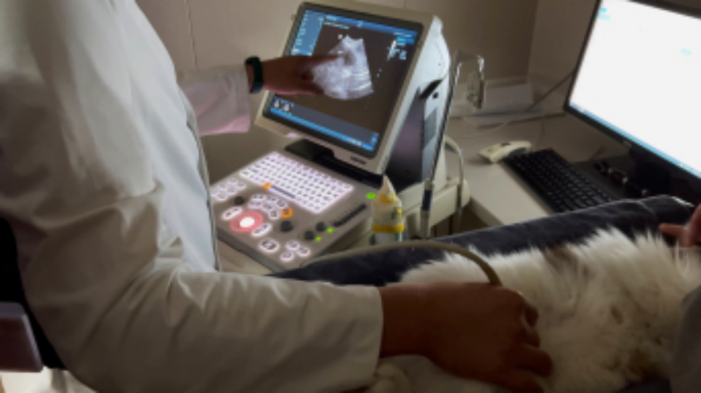
An ultrasound machine consists of:
The main unit (console)
The brain and core, responsible for signal transmission, reception, processing, calculation, and image generation.
The display
Displays the ultrasound image.
The core component - the probe (transducer)
Function: It acts as both a "speaker" to transmit ultrasound waves and a "microphone" to receive echoes.
Diversity: Probes vary in shape and frequency depending on the examination site:
▪ Convex array probe: Most widely used (e.g., abdominal and pelvic examinations).
▪ Linear array probe: Commonly used for superficial tissues (e.g., thyroid, breast, and blood vessels).
▪ Intracavitary probe: Such as transvaginal and transrectal probes, which provide a clearer view of pelvic organs.
▪ Phased array probe: Commonly used for cardiac examinations (e.g., cardiac color Doppler ultrasound).
Frequency selection: High-frequency probes offer high resolution (clearer details) but weaker penetration (suitable for superficial tissues); low-frequency probes offer greater penetration (suitable for deeper organs) but relatively lower resolution. Technicians must select the appropriate probe based on the examination target.

What can B-ultrasound see? Widely used in clinical practice!
Ultrasound examinations cover a wide range of areas, encompassing almost every system in the body (air-containing organs such as the lungs and intestines are significantly affected by air interference):
01 Abdomen: Liver, gallbladder, pancreas, spleen, and kidneys (assessing size, morphology, structure, and the presence of stones, cysts, and tumors).
02 Urinary system: Kidneys, ureters, and bladder (assessing stones, hydrops, tumors, and benign prostatic hyperplasia).
03 Gynecology: Uterine and ovarian size, position, and development; monitoring follicles; diagnosing fibroids, cysts, and ectopic pregnancies; and monitoring fetal development throughout pregnancy.
04 Obstetrics: Fetal screening (NT scan, major and minor fetal anomaly screening), assessment of placental location, and amniotic fluid volume.
05 Cardiac (echocardiogram): Assessment of cardiac structure (atrial and ventricular size, valve opening and closing), function (systolic and diastolic function, ejection fraction), and pericardial effusion (requiring specialized cardiac probes and more advanced equipment). 06 Superficial organs: Thyroid (nodules, enlargement), breast (lumps, cysts), superficial masses, lymph nodes.
07 Vascular: Combined with Doppler function, it can evaluate cervical vascular conditions (carotid artery plaques, stenosis), limb vascular conditions (arteriosclerosis, thrombosis, venous valve insufficiency), etc.
08 Musculoskeletal system: Tendons (tears, inflammation), ligaments, soft tissue injuries, joint effusions.
09 Interventional guidance: Real-time guidance for punctures, biopsies, catheter placement, and other procedures (such as liver biopsy, renal biopsy, and pleural/peritoneal effusion drainage).
What preparations are needed before the examination?
The requirements for different examination areas vary. It is crucial to follow your doctor's instructions for preparation, as this is crucial for image clarity:
Fasting
(Usually, fasting for 8 hours). Abdominal examinations (liver, gallbladder, pancreas, spleen, etc.) require fasting to reduce gastrointestinal gas interference. Avoid gas-producing foods 1-2 days before the examination. Holding Urine (Full Bladder)
Pelvic, urinary tract (bladder, prostate), and early obstetric examinations often require holding urine. This opens an acoustic window, pushing away intestinal gas and facilitating visualization of the uterus, adnexa, bladder wall, and underlying structures. The bladder can be emptied promptly after the examination.
No Special Preparation Required
Examinations of the thyroid, breast, extremity vessels, and superficial masses generally require no special preparation.
Clothing
Wear loose-fitting clothing that exposes the area being examined. Inform your doctor of your medical history, surgical procedures, and medications.
Interpreting Ultrasound Images
Ultrasound images are two-dimensional slices composed of varying grayscale levels. The physician/technologist will comprehensively assess the image based on characteristics such as the shape, size, boundaries, internal echogenicity (uniform/inhomogeneous, high/low/anechoic), echogenicity (e.g., duct-like, lattice-like), and the presence of posterior echogenicity enhancement/attenuation. Examination Report: A qualified sonographer (usually a physician) will provide a diagnosis based on the image and clinical data. The report describes the structural features and provides a diagnostic opinion (e.g., "Possible cyst considered," "Solid nodule, further examination recommended," etc.).
Is ultrasound safe?
1. No radiation: Ultrasound uses mechanical waves (ultrasound waves), not technologies like X-rays or radioisotopes that emit ionizing radiation.
2. Safe and reliable: Extensive medical research and long-term clinical practice have demonstrated that ultrasound energy used for medical diagnosis is safe and harmless to human tissue. Even for routine prenatal checkups, it is currently recognized as one of the safest imaging modalities.
3. Non-invasive and convenient: No injections are required (conventional ultrasound), the examination is relatively comfortable (except for the need to hold urine), and it can be repeated.
Note: While safe, ultrasound energy is not completely harmless. It should be used appropriately and as needed under the guidance of professional medical personnel.
New Developments and Future Directions in Ultrasound Technology
Ultrasound technology is rapidly evolving and is a key branch of medical imaging:
3D/4D ultrasound
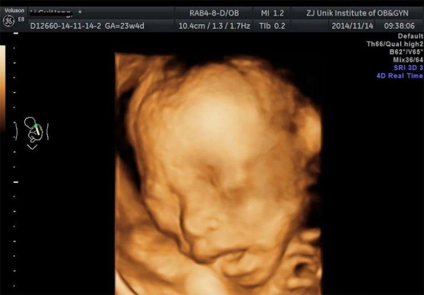
From 2D to 3D dynamic imaging (fetal facial imaging is the most surprising feature). Contrast-enhanced ultrasound
Intravenous injection of a special microbubble contrast agent significantly improves the detection and characterization of small lesions (especially in the liver).
Elastography
Assess tissue stiffness (e.g., the degree of liver fibrosis and the firmness of breast/thyroid nodules).
Interventional ultrasound
The application areas are continuously expanding, moving from diagnosis to precision treatment.
Artificial Intelligence (AI)
AI-assisted image interpretation, automated measurement, and assisted diagnosis of lesions will significantly improve diagnostic efficiency and accuracy. This will be one of the future battlegrounds for medical technology talent.
B-ultrasound Conclusion
B-ultrasound is like having a pair of non-invasive "sharp eyes," using ultrasound waves to penetrate human tissue and reveal its inner secrets. It is safe, convenient, and practical, making it an indispensable tool in clinical diagnosis. As a future force in medical technology, whether studying imaging technology, medical laboratory testing, or clinical medicine, a deep understanding of the principles and applications of B-ultrasound will lay a solid foundation for your future career.


