The widespread use of medical
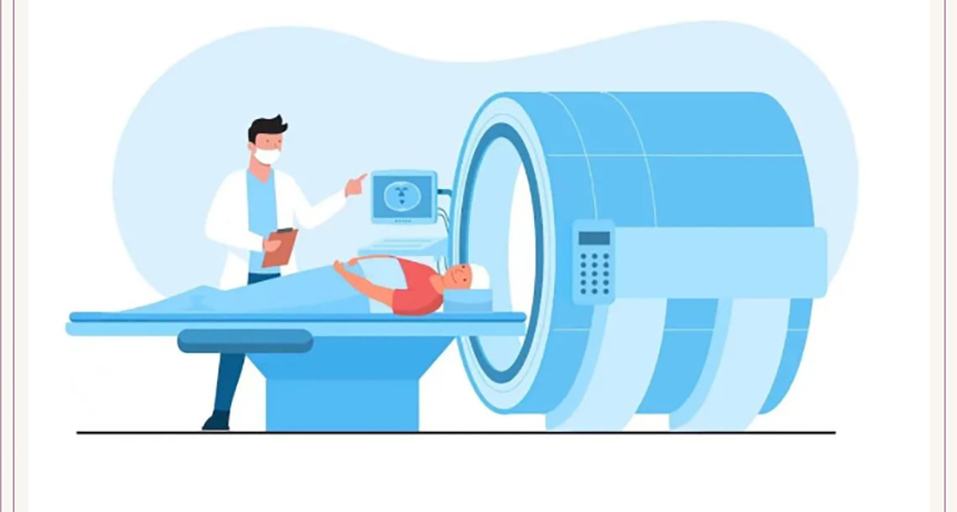
X-ray imaging examinations has made medical exposure the largest and ever-increasing source of artificial ionizing radiation to the public worldwide. X-ray diagnosis uses the principle that X-rays penetrate different tissues and organs of the human body differently to form images that match the density of the internal structure of the human body, thereby helping doctors diagnose the condition and improve the effectiveness of disease diagnosis and treatment. X-ray imaging diagnosis usually includes X-ray photography (such as radiography), X-ray fluoroscopy (such as chest X-ray, gastrointestinal angiography), computed X-ray tomography (CT), dental X-ray photography (such as dental X-ray, etc.) from relevant departments of the hospital. Panoramic machine, oral CT), mammography (such as mammography), bone density scan and interventional radiology procedures, etc.
What is the radiation dose received by the subject during radiological diagnosis?
The radiation dose to a patient during an X-ray examination procedure depends on factors such as the type of equipment, type of examination procedure, patient size and body part examined, and exposure parameters.
The effective dose (in millisieverts, mSv) is commonly used to compare patient doses in different X-ray diagnostic procedures. The radiation dose from conventional X-ray photography is relatively low, while the radiation dose from CT scan is higher. For example, for the same chest examination, the dose of a chest X-ray examination is about 0.02-0.1 mSv, while the dose of a CT chest scan is about 6-8 mSv, which is dozens to hundreds of times that of a chest X-ray; the dose of a low-dose chest CT scan is It is about 1.5mSv, which can significantly reduce the patient dose. In medical exposure, the contribution of CT to the collective dose of the population has reached more than 60%.
Dangers of X-rays
The effects of X-rays on the human body are mainly divided into harmful tissue reactions (previously called deterministic effects) and stochastic effects.
There is a threshold dose for deterministic effects. When the threshold is exceeded, there is a certain probability that harmful effects will occur. The higher the dose, the greater the severity of the effect. When the dose is below the threshold, no harmful effects will occur. Stochastic effects mainly include carcinogenic effects and genetic effects. There is no dose threshold for stochastic effects, which is characterized by the probability of occurrence being proportional to the dose, but the severity being independent of dose.
In the field of X-ray diagnosis, the patient dose in a single Multiple or prolonged exposures may cause deterministic effects such as skin erythema, hair loss, burning sensation, edema, stinging, and even necrosis and ulcers.
How to protect sensitive groups
Infants, children, pregnant women, women of childbearing age and other groups are susceptible to radiation damage.
The lifetime risk of irradiated children developing solid cancers other than leukemia is 2 to 3 times that of the general population, and the risk of leukemia is much higher. A growing number of epidemiological studies have shown that CT examinations in children are associated with an increased risk of brain tumors and leukemia. Therefore, the legitimacy of children’s diagnostic medical exposure (especially CT examinations) should be strictly judged, unnecessary CT examinations should be avoided, and the exposure parameters should be optimized to reduce the exposure dose to children.
With regard to X-ray examinations for pregnant women and women of childbearing age, radiological examinations that would cause exposure to the abdomen or pelvis of women who are pregnant or may become pregnant may be performed only if there is a clinically sound reason to do so, and otherwise such exposure should be avoided.
The lower abdominal examination of women of childbearing age should be performed within ten days after menstruation; the risk of radiation is greatest during the fetal organ formation period and early fetal period during the entire pregnancy, and the fetal central nervous system is most sensitive to radiation between 8 and 15 weeks after fertilization. , followed by the next. Women of childbearing age who are 8 to 15 weeks pregnant are not allowed to undergo lower abdominal X-ray examination unless there are special needs.
When conducting X-ray examinations on sensitive groups, the radiation field should be strictly limited and non-examination parts should be protected. In particular, the shielding and protection of gonads, eye lenses, and children's bones should be strengthened. Optimize irradiation parameters to achieve clinical diagnosis purposes with a reasonable and lowest possible dose.
How to reduce the harm of X-rays
Under the premise of legitimacy, the benefits of clinically appropriate X-ray imaging far outweigh the risks. The absolute risk to individual patients is small, but the risk of random effects on the population cannot be ignored. Therefore, low-dose examination procedures or alternative procedures that do not involve ionizing radiation should be considered clinically as appropriate, and X-ray examinations that do not comply with the principle of legitimacy should not be performed. Minimize the risk from radiation hazards by reducing unnecessary exposure to ionizing radiation.
When it is clinically necessary to use X-ray examination to confirm the diagnosis, while satisfying the clinical diagnosis, a lower-dose X-ray examination procedure should be used as much as possible, and always follow the as low as reasonably achievable (ALARA) principle, and Repeated inspections should be avoided.
Before subjects undergo X-ray examinations, medical institutions should strictly control the scope of the radiation field and equip the subjects with necessary radiation protection equipment to protect sensitive organs or tissues adjacent to the radiation field (such as gonads, eye lenses, breasts, and thyroid glands). ) Take necessary shielding and protection measures to avoid direct exposure to the main X-ray beam and minimize the patient’s radiation exposure. It is recommended to use wrapped shielding protection measures for subjects during CT scans.
How diagnostic imaging and radiology workers can avoid X-ray damage
Since the discovery of X-rays by Roentgen in 1895 and its clinical application, it has been observed in many countries, including my country, that radiologists and technicians who were engaged in clinical radiological imaging diagnostic examinations in their early years have a significantly increased risk of skin cancer, leukemia and other tumors. With the advancement of science and technology, especially the widespread adoption of compartmental operations, the exposure dose to imaging diagnostic radiation workers has been significantly reduced. However, in recent years, radiation health monitoring work has found that the annual dose of interventional radiation workers is relatively high, and the risk of radiation diseases such as peripheral blood lymphocyte chromosome aberration rate and eye lens opacity has increased significantly.
To protect personnel engaged in diagnostic imaging and radiology, we must first ensure that equipment and facility performance testing and protection monitoring meet the requirements of relevant national regulations and standards. It is very safe for medical staff operating in the outer compartment of the computer room that has passed the protection test. The radiation leakage they receive is extremely low, and there is no need to worry about radiation hazards. For interventional professionals and other personnel operating in the room, they need to protect radiation-sensitive organs such as the eye lens, thyroid gland, gonads, etc. by wearing personal protective equipment.
When operating the equipment, the staff should control the irradiation field as much as possible and optimize the irradiation conditions without affecting the purpose of irradiation, so as to reduce the radiation dose of the main ray and at the same time reduce the risk of radiation exposure of the staff.
Finally, radiation workers must receive regular radiation protection training and occupational health examinations, and wear personal dosimeters in their daily work to monitor whether the amount of radiation received by the whole body, eye lens, hands and feet exceeds the standard.