As medical technology continues to advance and people pay more attention to health, radiology technology plays an increasingly important role in medical diagnosis and treatment. In modern hospitals, it is crucial to build an efficient and safe radiology department. However, this involves many complex factors that require comprehensive consideration and reasonable planning. To this end, we specially invited medical facility planning experts to explain the key points that need to be paid attention to when building a radiology department, so as to help medical institutions better construct and operate radiology departments.
Safety and Radiation Protection
Radiology departments involve the use of radiation and radioactive sources, so safety and radiation protection are one of the primary considerations. We need to ensure that facilities comply with relevant safety standards and regulations and take effective radiation protection measures to protect the health and safety of medical staff and patients. This includes the use of protective equipment that meets standards, regular radiation dose monitoring and training of medical staff in the correct use of protective measures.
Device selection and configuration
Building a radiology department requires selecting appropriate radiology equipment based on the size and needs of the hospital. Selecting appropriate equipment, including CT, X-ray, MRI, etc., requires full consideration of factors such as the hospital's service scope, patient population, and medical level. Properly configuring the location and layout of radiation equipment can not only improve the efficiency of equipment use, but also meet the needs of medical work and ensure the smooth progress of medical services.
1. x-ray
First, X-ray examination is the most common imaging examination and the most commonly used examination method. When a patient is clinically suspected to have cough, fever, chest pain, or chest tightness, an X-ray examination will be ordered first to preliminarily rule out lung lesions and see if there is pneumonia or space-occupying lungs.
Second, when trauma occurs, X-ray examination is most commonly used. For example, if there is trauma to joints such as hands and feet, after X-rays are taken, a preliminary diagnosis can be made to determine whether there is a fracture or dislocation.
Third, X-ray examination is still often used as a routine item to diagnose cervical and lumbar degeneration or joint degeneration.
2. CT: CT is used to observe lesions in the brain and chest. When space-occupying lesions are found, enhanced scans are performed, which is helpful for qualitative examination of the lesions. For abdominal lesions, CT-enhanced examination is generally performed directly, which is helpful to detect the lesions. CT is also called computerized tomography. It uses accurately collimated x-ray beams, gamma rays, ultrasound, etc., together with extremely sensitive detectors to perform cross-sectional scans one after another around a certain part of the human body. According to the scan time Fast, clear images and other features. It can be used to examine a variety of diseases; depending on the type of radiation used,
ct can be divided into the following types:
The first type is x-ray CT.
The second type is ultrasound CT and gamma ray CT, etc.
3. MRI: MRI examinations have many purposes and can be used to detect problems such as tumors, bleeding, injuries, vascular diseases, or infections. For example: head. MRI can check the brain for tumors, aneurysms, brain bleeds, nerve damage and other problems such as damage from a stroke. Problems with the eyes and optic nerve, ears and auditory nerve can also be found. Chest. A chest MRI can look at the heart, valves, and coronary blood vessels and can show whether the heart or lungs are damaged. It can also be used to look for breast cancer. Blood vessel. The use of MRI to look at blood vessels and the blood flow through them is called magnetic resonance angiography (MRA). Problems with the arteries and veins, such as aneurysms, blocked blood vessels, or tears in the lining of blood vessels (dissection), can also be found. The blood vessels can be seen more clearly with the contrast agent. Abdomen and pelvis. MRI can detect problems with abdominal organs and structures, such as the liver, gallbladder, pancreas, kidneys, and bladder, and can detect tumors, bleeding, infections, and blockages. The uterus and ovaries in women and the prostate in men can be observed. Bones and joints. MRI can check for bone and joint problems such as arthritis, TMJ problems, bone marrow problems, bone tumors, cartilage problems, torn ligaments or tendons, or infections. MRI can also be used to diagnose fractures when X-ray results are unclear. MRI is more common than other tests when checking for some bone and joint problems. spine. MRI can check the discs and spinal nerves for conditions such as spinal stenosis, disc herniation, and spinal tumors.
Angiography: mainly to display the characteristics of lesions more clearly. Commonly used angiography examinations for the cardiovascular system, including coronary angiography, can determine whether there is stenosis of the coronary arteries, the location and degree of stenosis, and coronary angiography, which is used to diagnose coronary heart disease. It is the gold standard and can be used to guide interventional diagnosis and treatment of coronary heart disease or surgical bypass surgery. In addition, angiographic examinations of the cardiovascular system also include pulmonary angiography, which can determine whether there is pulmonary embolism. If necessary, interventional procedures such as pulmonary artery balloon dilatation can be performed. In addition, angiography of the cardiac chambers can be performed to evaluate cardiac function. It is the gold standard for functional diagnosis and can also determine whether there are ventricular aneurysms, cardiac enlargement and other related conditions.
Space planning and environmental design
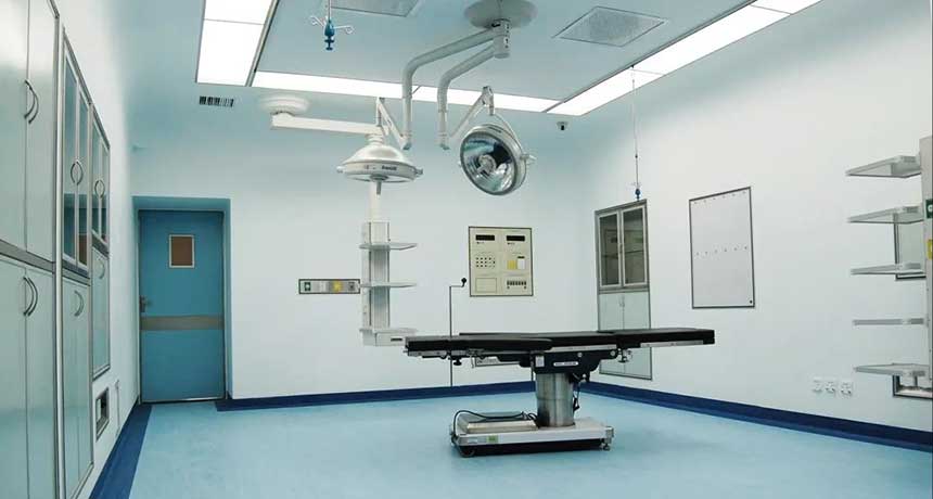
The space planning and environmental design of the radiology department directly affect the efficiency and quality of medical work. We need to reasonably plan the layout and space utilization of the radiology department based on the needs of the radiology equipment, ensure that the distance between the equipment and the width of the passage meet the requirements, while ensuring the comfort and ventilation of the environment. In addition, aspects such as radiation protection and exhaust systems of radiological equipment need to be considered to ensure the safety and comfort of medical staff and patients. When doing radiation protection construction in the radiology department of a new hospital, you must choose a good radiation-proof lead door. When the radiology department is doing radiation protection construction, you must not only choose a good radiation-proof lead door, but also choose a construction team to carry out radiation protection construction for the radiology department computer room. , everyone knows that there are many radiological equipment in the radiology department of the hospital, which will produce ionizing radiation when scanning and testing patients. This ionizing radiation is very helpful in detecting the cause of the patient's disease, but it is very difficult for the medical staff who have been in the radiology department for a long time. Harmful. Long-term radiation damage to the body can lead to cancer in some organs of the body. When choosing a radiation-proof lead door, you need to know which computer room it is used for in the radiology department. For example, a CT computer room needs to purchase 4 lead-equivalent radiation-proof lead doors. A room with limited ability to shield the ionizing radiation in the computer room.
Personnel training and technical support
The operation of a radiology department requires professional medical staff and technical support teams. Before setting up a radiology department, we need to make preparations for training and technical support for medical staff to ensure that they have the necessary skills and knowledge to operate radiation equipment proficiently and handle related problems. At the same time, it is also necessary to establish a complete technical support system to solve equipment failures and technical problems in a timely manner to ensure the smooth progress of medical work.
Legal, regulatory and regulatory requirements
The radiology department involves the use of radiation and radioactive sources, and therefore needs to strictly comply with relevant laws, regulations and regulatory requirements. Before building a radiology department, we need to understand and comply with relevant national and local laws and regulations to ensure that the construction and operation of the radiology department comply with legal requirements and pass the approval and supervision of relevant departments. In addition, it is necessary to establish a sound quality management system, conduct regular quality assessments and supervision inspections, and improve the management level and service quality of radiology departments.
expert's point:
"The radiology department is an important medical department in the hospital. Building a radiology department requires comprehensive consideration of safety, equipment selection, space planning and other factors. We hope that medical institutions can take the construction of radiology departments seriously to ensure the smooth progress of medical work. , to provide patients with better medical services."
Through the interpretation of the above key points, we have an in-depth understanding of the important aspects that need to be paid attention to when building a radiology department. This will help medical institutions better plan and build radiology departments, improve medical service levels, and provide patients with better medical services.


