Electrocardiography is a subject that diagnoses through graphics, and the quality of graphics is the basis for correct diagnosis of
ECG.
This article will summarize the operating techniques of electrocardiogram. By standardizing pre-operation preparations, potential placement requirements, use of additional leads and handling of special patients, we will summarize and improve electrocardiogram operation skills to obtain higher quality electrocardiograms.
1. Nursing
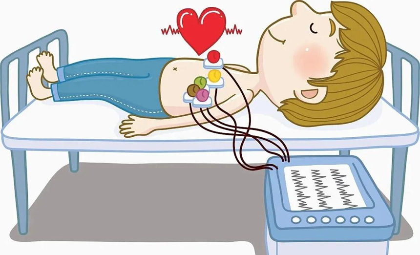
Preparation before operation: The environment requires a suitable room temperature, maintained at 18°C ~ 26°C, to avoid electromyographic interference caused by cold. Make sure the patient can lie down comfortably with his limbs relaxed. If the examination bed is too narrow, place the patient's hands under the buttocks to avoid arm muscle tension.
Instrument requirements: The electrocardiograph is a measuring device and requires annual measurement and testing to ensure the authenticity of the recorded data. If the lead wires are damaged or aged, replace them promptly. Before use, check whether each lead wire is connected correctly and whether it is loose. During operation, do not twist the lead wires and keep them as far away from power sources and other electrical appliances as possible to avoid interference from alternating current and electromagnetic waves. Arrange them promptly after use and place them properly.
The patient requires that the patient's privacy be maintained. If the skin where the electrode is placed is dirty or has excessive hair, it should be cleaned or shaved first. During the examination, the baby should be placed in a supine position and kept quiet. If the baby is crying and uncooperative, the baby can be sedated in advance or be asleep before the examination. Infants and young children have small chests, narrow intercostal spaces, and thin skin. Use disposable electrodes as much as possible.
2. Placement of electrodes
Limb leads ECG limb leads are clamped on the left wrist, right wrist, left ankle, and right ankle respectively. They should not be placed on the arms or calves. They should be placed on a flat and fleshy part. Limb lead electrodes are placed equidistant from the heart, placing the electrodes in the same location on each limb. If the limb part cannot be placed (such as amputation, injury), please place the electrode closer to the torso.
Chest leads V4, V5, and V6 should be at the same level. If the position of V5 is difficult to determine, place V4 and V6 in the middle of the same level; V7, V8, and V9 should also be at the same level as V4. Female patients with breast sagging should hold up their breasts and place the V3 and V4 electrodes on the chest wall at the lower edge of the breast. They should not be placed on the breast. Those who have mastectomy should be noted. When tracing V7, V8, and V9, the patient must be in the supine position and should not be traced while lying on the side or in other positions. Do not place the electrodes of the left and right lower limbs on one side of the lower limb. The synchronous 12-lead electrocardiograph is equipped with a right lower limb reverse drive circuit, which can effectively suppress AC interference. If placed on the same side, the anti-AC interference performance will be reduced.
3. Infant leads
In infants and young children under 3 years old, the right ventricle plays a dominant role, so lead V4R should be added during tracing. If a 12-lead electrocardiograph is used, the V3 electrode is often placed at the V4R position to record the current spreading in the right chest of the heart. In some children with congenital heart disease, blood may be shunted to the right, causing changes in the right ventricle. At this time, lead V4R should also be added.
Adding 18 leads. Currently, conventional 12-lead electrocardiograms are mostly used in clinical practice. However, 12-lead electrocardiograms can only accurately locate lesions on the anterior wall, anteroseptal wall and inferior wall of the heart, but cannot detect lesions and electrical activity on the posterior wall and right ventricle. It cannot be recorded, so for patients with chest pain or patients with acute myocardial infarction, leads V3R, V4R, V5R, V7, V7, and V9 should be added to make up for this deficiency. Studies have found that adding right ventricular and posterior wall leads to the conventional 12 leads in acute chest pain can increase the detection rate of ST segment elevation by 12%.
The 18 leads can also more comprehensively reflect the changes in the ST segment, Q wave and T wave of each link, and can predict vascular lesions earlier, providing a basis for early clinical treatment. If the R wave increases in V1 and V2 and appears Rs-shaped, it may indicate the possibility of myocardial infarction in the posterior wall. An 18-lead electrocardiogram is added to prevent missed diagnosis of right ventricular and posterior wall myocardial infarction and improve the positive detection rate of coronary heart disease.
Identify dextrocardia
When the P waves in lead I of the electrocardiogram are all downward, the P waves in lead avR are upright to rule out misconnection between the left and right hands, and the R waves in the chest leads are getting smaller and smaller, and when dextrocardia is suspected, limb leads and limb leads after reverse connection of the upper limbs should be added. V3R, V4R, V5R. If the ECG shows all normal lead shapes at this time, it indicates that the patient has mirror dextrocardia.
Use of additional leads
When V1 and V2 are QS-shaped when the next intercostal space is added, the diagnosis of myocardial infarction should not be made casually. It must be combined with the patient's age, medical history, and symptoms. If the patient has no history of coronary heart disease and no symptoms of chest pain or chest tightness, it is possible that the electrocardiogram is clockwise transposed. At this time, record the next rib or two intercostals at the original position where leads V1 and V2 are normally recorded. If there is r It is normal to have an embryo.
When it is necessary to differentiate between Brugada waves in the last intercostal space and right bundle branch block, the original normal recording position of V1 and V2 leads ECG can be raised by one intercostal space or two intercostal spaces and then recorded.
If it is a Brugada wave, the V1 or V2 lead located in the higher intercostal space records a more typical and diagnostic Brugada; because most (more than 80%) of the abnormal electrical activity of the Brugada wave is limited to the right ventricular outflow tract, it is very limited. parts. In right bundle branch block, V1 is mainly rsR type, the terminal S wave of QRS wave in leads V5 and V6 is widened, but the terminal S wave of Brugada wave in lead V5 is not widened.
Adding lead S5 When encountering complex electrocardiograms where the P wave is difficult to identify, you must try to find the P wave. At this time, lead S5 can be added. Lead S5 is a bipolar lead, and the P wave is clearer in this lead. The right-hand electrode (negative electrode) is placed on the sternal manubrium, and the left-hand electrode (positive electrode) is placed on the fifth intercostal space at the right edge of the sternum. At this time, the recording is an I-lead electrocardiogram. This method enables a high P wave detection rate and is used to analyze cardiac arrhythmias.
Dynamic observation and clear diagnosis
Slowing down the ventricular rate, clarifying the atrioventricular relationship or changing the conduction ratio to confirm the diagnosis. Breath-hold test, pressing the carotid sinus, and pressing the eye frame are all commonly used methods.
When atrial flutter, sinus tachycardia, and atrial tachycardia cannot be diagnosed, breath holding can be used to slow down the ventricular rate and reveal the hidden P wave or FL wave. Suspected supraventricular tachycardia can be terminated by breath-holding and carotid sinus compression. Suspicious atrial flutter and atrial tachycardia can be diagnosed clearly by changing the conduction ratio through breath holding.
Increasing the ventricular rate is also a method of identification in our work. Early repolarization is a normal variation of the electrocardiogram, which is manifested as ST segment elevation behind the J point. In order to identify that the elevation of the ST segment is just a normal variation of the electrocardiogram, we will advise such patients to run or walk quickly, increase the heart rate and then record the electrocardiogram again to observe the changes in the ST segment. If it returns to normal, it is a normal phenomenon. .
Dynamic observation: For patients with chest pain and suspected coronary heart disease, do not make a diagnosis lightly when there is no change in the electrocardiogram. Because some patients may show pseudo-improvement in their ECG at this time, the patient can be asked to do it again later, or the patient's previous ECG can be reviewed to dynamically observe changes in the ECG.
Summarize:
In the practice of electrocardiogram examination, using the techniques summarized above can greatly improve the diagnostic accuracy of electrocardiogram. Adding 18 leads to elderly patients and patients with acute chest pain can improve the detection rate of latent coronary heart disease and patients with mild lesions and good collateral circulation. With the development of congenital heart disease, valvular disease and complex cardiac surgery, some arrhythmias have become more complex, and the use of some additional leads can clarify the diagnosis of arrhythmia. For example, the use of S5 lead can significantly improve the diagnosis of complex atrial arrhythmias, which is consistent with the report of Chen Xiuling et al. During the physical examination, some abnormal electrocardiograms must be carefully identified. For example, adding the next intercostal space can eliminate abnormal Q waves caused by the position of the heart and avoid misdiagnosis of anterior septal myocardial infarction. To sum up, although electrocardiogram is a subject that diagnoses through graphics, we must also combine it with clinical practice to ensure the authenticity of the obtained graphics and identify the graphics, so that we can make a more accurate interpretation of the electrocardiogram and provide clinical A more powerful basis for diagnosis.