I. Technical Specifications
1. Combined High frequency & High voltage generating system
1.1 Tube Focus: 0.6mm/1.8mm
1.2 Anode thermal capacity: 35kJ(47kHu)
1.3 Tube thermal capacity: 650kJ(867kHu)
1.4 Output power: 5kW
1.5 Inverter frequency: ≥40kHz
1.6 Continuous fluoroscopy (Manual & automatic mode)
1.6.1 Tube voltage: 40kV~120kV
1.6.2 Tube current: 0.3mA~4mA
1.6.3 Automatic brightness tracking function
1.7 Pulse fluoroscopy
1.7.1 Tube voltage: 40kV~120kV
1.7.2 Tube current: 0.3mA~30mA
1.7.3 Pulse frequency: intelligent-control, can reduce X-ray radiation, enhance single frame image quality, and increase continuous work time.
1.8 Photography Mode
1.8.1 Photography tube voltage: 40kV~120kV
1.8.2 Photography tube current: 25mA~100mA
1.8.3 Photography mAs: 1mAs~180mAs
1.9 Collimator: electrical and rotating linear symmetry
1.10 Working Environment Conditions
1.10.1 Environment temperature: 10°C—40°C
1.10.2 Relative humidity: 30%—75%
1.10.3 Atmospheric pressure: 700hpa—1060hpa
1.11 Working Power Supply
1.11.1 Power supply voltage: Single phase, 220V±22V
1.11.2 Power supply frequency: 50Hz±1Hz
1.11.3 Internal resistance: no more than 1Ω
2. Imaging System
2.1 Imaging intensifier:Imported Canon 9″ E5764SD-P3, three-field visual (4.5”/6”/9”)
2.2 Camera:Ultra low-light CCD camera, one mega pixels with progressive scanning by black and white, 1024x1024
2.3 LCD display: 24 inches, resolution is 1920x1200, 19inch 1280*1024, frequency is 60Hz
2.4 Imaging Acquisition and Processing Workstation
2.4.1 Register: register & save, medical records checking, Worklist
2.4.2 Acquisition: image collection, ready to video, reset, horizontal & vertical mirror, window modulation, magnifier, negative image, open silhouette, edge enhancement, recursive noise reduction.
2.4.3 Processing: four-window, nine-window, sharpening, mirror horizontally and vertically, image text, image length measurement.
2.4.4 Report: save, preview, expert template
2.4.5 Dicom function:Dicom browse, web service.
2.5 Image sharpness index
2.5.1 Gray scale:≥ 11 (Inspection process requirement)
2.5.2 Lines of resolution:≥ 2.0 LP/mm
3. Mechanical Performance
3.1 Backward & forward movement: 200mm
3.2 Rotation around horizontal axis: ±180°
3.3 Rotation around vertical axis: ±15°
3.4 SID: 1000mm
3.5 C-arm open: 760mm
3.6 C-arm depth: 670mm
3.7 Slide along the obit: 120°(+90°~-30°)
3.8 Pillar up & down movement: 400mm
3.9 Directive wheel and main wheel movement: directive wheel can move in any directions, main wheel can rotate from -90°to + 90°
3.10 Electric support arm
3.11 Full-balance: When the equipment is under the condition of unlocked mechanical motion, C arm can keep the balance in any position and angle, do not slip.
3.12 Light thrust
II. Standard Configuration
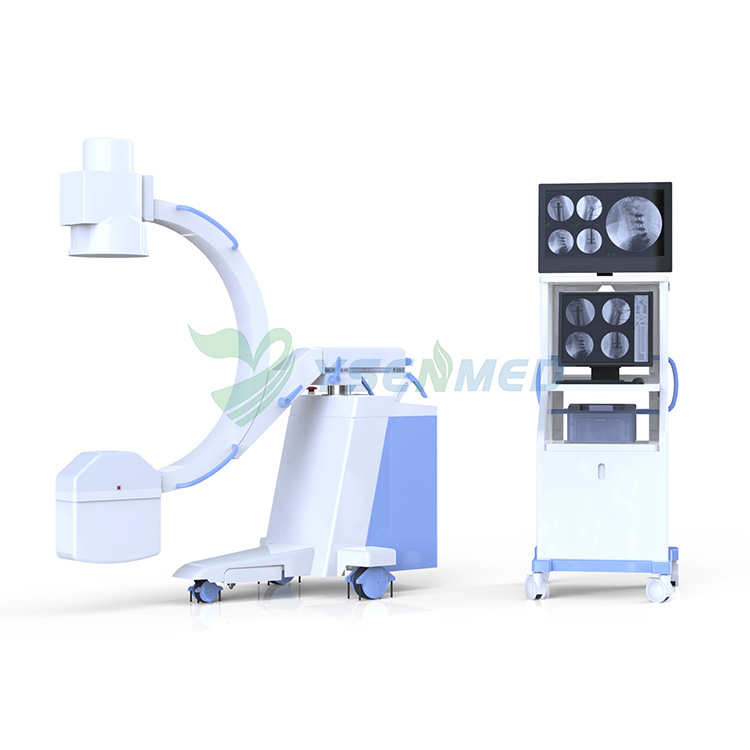
1. Main frame of C-arm 1 set
2. High-frequency high-voltage X-ray generator and
high frequency inverted power supply ( 5kW、40kHZ、120kV) 1 set
3. 19 inch LCD display, resolution 1280*1024 1 set
4. 24 inch LCD display, resolution 1920x1200 1 set
5. 9 inch Canon imaging intensifier, three filed 1 set
6. Medical use one mega pixel digital camera with ultra low light ( 1024x1024, 12bit, SNR≥64) 1 set
7. Digital acquisition and processing workstation 1 set
8. Mini groove grid 1 set
9. Electric adjustable collimator 1 set
10. Hand controller 1 set
11. The Red Cross locator 1 set
III. Features
- High quality combined high-frequency and high-voltage X-ray generator can reduce X-ray radiation, save and efficient.
- kV、mA automatically tracking function makes the brightness and sharpness of image in the best condition.
- Real-time and continuous fluoroscopy mode reduces the X-ray dose and ensures superior image quality, which can meet the demand of high-precision and more complicated minimally invasive surgery, and also can effectively protect the security of medical staff and patients。
- Adopting operating interface with human-graphic LCD touch- screen, which makes the operation easier and more convenient.
- Hand controller design, easy to operate.
- Canon brand imaging intensifier with reliable and stable quality, which ensures high definition image.
- Ultra low light i mega pixel camera, provide sharpen image.
- Standard configuration workstation with advanced image processing software, which makes the good image and easy to operate and diagnose. Dicom 3.0 interface can be connected with HIS, RIS and PACS in the hospital improve work efficiency.
- Electric assist support arm design, safe use.
- Beautiful out appearance design, compact structure and new frame design.
- Realize digital radiography and fluoroscopy, makes the surgery more safe and convenient.
- Popular in both domestic market and overseas market because of the good quality and design.