Professional and accurate, delicate and comprehensive:
High-Definition.
Super-fast Workflow.
Low-Dose.
Long Service Life.
Advanced Aplication.
A high-end 32-slices CT with Optimal Resolution:
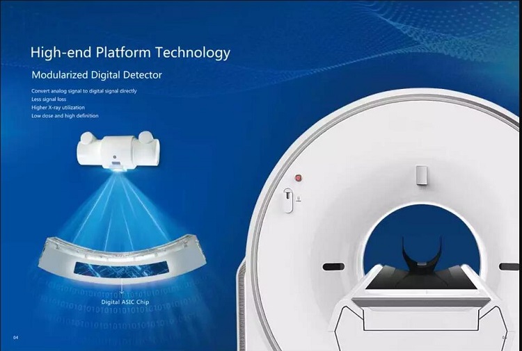
ScintyStar Detector:
– Owning the Intellectual Property Rights..
– New Modular Integrated Detector.
– High Contrast Resolution MTF 10%; 13 lp/cm.
– Ultra-high speed rare earth scintillator material. – This material increases the quantum detection efficiency, and has a very fast decay time, thus can improve the spatial resolution and produce good image quality evan at a lower dose.
– ASG + ASIC design for maximum signal-to-noice ratio. – The detector module design is fully integrated and miniaturized to meet important performance parameters: low scatter, low electronics noice, high signal-to-noice ratio.
Low Dose Technology:
– imA (intelligent mA). – The output milliamperes of the X-Ray tube are automatically controlled according to the size of the patients and the scanning position, so as to ensure a more balanced image at each layer, while the patient receives a lower radiation dose.
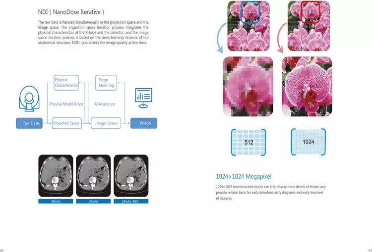
– NDI (NanoDose Iterative). – The raw data is iterated simultaneously in the projection space and the image space. The projection space iteration process integrates the physical characteristics of the X-Ray tube and the detector, and the image space iteration process is based on the deep learning network of the anatomical structure. NDI+ guarantees the image quality at low dose.

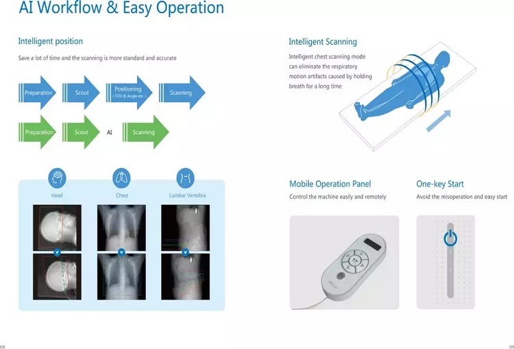
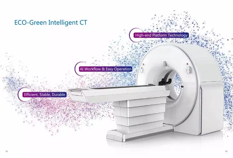
Super-fast Workflow. One-key Intelligent Scanning.
– Large Pitch Spiral Scanning with SAC Technology.

High-speed Recontruction System

Optima Design:
– One Side Integrated Control. – Optimize System Control Layout. Improve Systematic Process Flow. Ensure Product Quality and Stability. Improve After-sales Maintenance Efficiency.
– Thermal Insulation Design. – Improve Heat Dissipation Efficiency. Extend the Life of Detector. Ensure the Image Quality.
– The Integrated Casting of Stator and Rotor. – Min Vibration During Rotation. Min Deformation During Rotation.
– High Precision Bearing. – Zero Error and Zero Runout under High Speed Rotation. Achieve Military and Aerospace Level Requirements. Long Service Life and Excellent Stability.
– Multi-point Temperature Control Technology. – Automatically Monitor the Temperature. Ensure the Stability of System Operation.
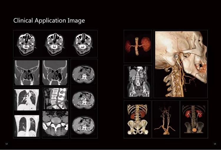
Clinical Application Image

Cloud diagnosis. – Famous radiologists diagnose through remote image diagnosis solution, improving primary hospital diagnosis ability.
Cloud storage. – MinFound Cloud storage is safe, stable and able to save much cost: payable based on requirement; it saves equipment purchasing and operation cost.
Global After-sales Cervice.
– Automatic Fault Warning Function.- Remote Service Function.
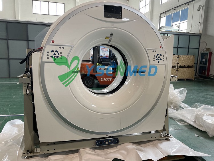
Gantry:
Aperture:70 cm.
Manual control from POD at console.
Gantry cooling: air.
Gantry rotation: belt-driven.
Slip ring type: low voltage.
Positioning system: 3D lasers.
HV Generator:
Power rating: 32 kW.
mA range: 10 – 300 mA.
kV range: 70 kV, 80 kV, 100 kV, 110 kV, 120 kV, 140
X-Ray tube:
Anode heat storage capacity: 5 MHU.
Anode heat dissipation: 735 KHU / мин.
Small focal spot size: 0.7 mm × 0.8
Large focal spot size: 1.2 mm × 1.4
Detector:
Number of detector rows: 16.
Number of detectors per row: 704.
Table:
Maximum motion range:1600 mm.
speed in horizontal direction:150 mm/s.
Motion tolerance in horizontal direction (200 kg):±1mm.
Table elevation range:440
Max weight load:450 lb (205 kg).
Foot pedal control, table horizontal motion.
Console:
Uninterrupted power supply UPS (3 KVA) to prevent sudden power outages and ensure the system stability.
Memory: 32 GВ.
Hard disk: 2TB + 1TB.
Frequency: 3.6 GHz.
Liquid crystal display of high resolution, displaying matrix (M * M): 1920 × 1200.
Progressive scan display.
DICOM 3.0 network interface.
Burn mode:
DICOM 3.0 print interface.
DICOM 3.0 output and input interface.
Auto-film.
Auto-voice and integrated intercom for communication between operator and patient.
Include DICOM PRINT,DICOM STORE,DIOCM QUERY,DICOM
RETRIVE,
Include HIS & RIS interface.
Raw data can be reconstructed on the workstation.
Scan and Image Reconstruction:
The fast rotation time: 0.75 s / 360°.
Longest scanning time:100 s.
Longest scanning range: 1200 mm on the original tabletop with head holder; 1300 mm on the original tabletop with the short extendet tabletop; 1400 mm on the original tabletop with the long extended tabletop.
Scout scan direction: anteroposterior, lateral.
Pitch range: Standard 0.25 – 1.75. Optional: 0.1 – 2.0
Max number of slices per rotation:16 / 32 slices.
Thinnest image slice thickness in 16-slice mode:25 mm.
Thinnest image slice thickness in 32-slice mode:25 mm.
Thinnest reconstructed slice thickness in 16-slice mode:25 mm.
Thinnest reconstructed slice thickness in 32-slice mode:25 mm.
Nominal slice thickness for axial scan: 1.25 mm,5 mm, 5 mm,10 mm.
Nominal slice thickness for helical scan: 1.25 mm,5 mm,3.75 mm, 5.0 mm,7.5 mm,10 mm.
Parallel image processing.
Simultaneous Reconstruction: Can reconstruct and reorganize the images simultaneously with a variety of options. Different reconstruction options are available within the scan protocol.
Scanning FOV:
Image reconstruction matrix:1024 × 1024.
Image display matrix:1024 × 1024.
PDU:
Input voltage: 3-phase 380 V, 50 Hz.
Power: 50 kVA.
System Software:
Beam hardening artifact correction software.
Posterior cranial fossa optimization.
Motion correction reduces motion artifact.
Pediatric protocol.
NDI (NanoDose Iteration).
Filming print software.
Remote maintenance system.
Accessory KIT:
Patient Table Pad, Head Holder, Head Holder Cushion, Phantom.
Clinical Application:
Include Volume Reconstruction (VR), Multi-planar Reconstruction
(MPR), Curved Planar Reconstruction (CPR), Surface Shaded Display
(SSD), MIP,MinP.
Option:
UPS.
Printer.
High Pressure Injector.
FOV: 500 mm.
Workstation:
– 16GB Memory, 24”color display, 1TB SATA hard disc.
– 3D image reconstruction: Include VR, MPR, CPR, SSD, Simulated
scalpel, Virtual endoscope, CTA remove bone, CTA subtraction.
– Cerebral hemorrhage measuring tool.
– Skeleton internal fixation fluoroscopy technique.
– Advanced automatic melting of bone.
– CT vessel analysis.
– Bone fragment removal.
– ROI creator.
– Stent planning.
Abdominal fat analysis.
The orthopaedic module.