X-ray machines can be divided into two uses: diagnostic and therapeutic
1. X-ray diagnosis
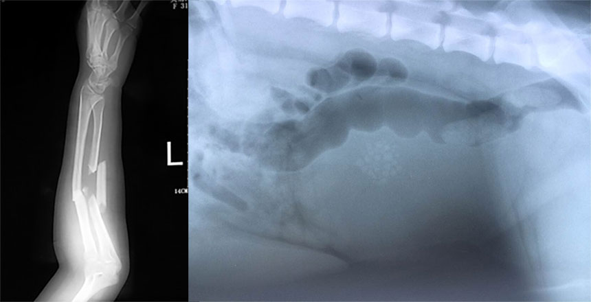
X-rays are used in medical diagnosis, mainly based on the penetration, differential absorption, photosensitivity and fluorescence effects of X-rays. Because X-rays are absorbed to varying degrees when passing through the human body. For example, the amount of X-rays absorbed by bones is greater than the amount absorbed by muscles. Then the amount of X-rays passing through the human body is different, which carries the density distribution of various parts of the human body. Information, there is a big difference in the intensity of fluorescence or photosensitivity caused on the fluorescent screen or photographic film, so shadows of different densities will be displayed on the fluorescent screen or photographic film (after development and fixation). Based on the contrast of shadow shades, combined with clinical manifestations, laboratory results and pathological diagnosis, it can be judged whether a certain part of the human body is normal. As a result, X-ray diagnostic technology became the world's earliest non-invasive internal organ examination technology.
X-ray examinations commonly used for the nervous system include skull radiography, cerebral angiography, CT, myelography, etc.; X-ray examinations commonly used for the circulatory system include cardiac fluoroscopy, cardiac telephotography, and cardiovascular angiography; X-rays commonly used for the digestive system Radiographic examinations include gastrointestinal tract imaging, X-ray pictures and imaging of the biliary system, CT examination of the liver, B-ultrasound, CT or angiography of the pancreas; X-ray examinations commonly used in the urinary system include X-ray abdominal plain films and intravenous urography , retrograde pyelography, renal angiography and CT; commonly used X-ray examinations of the sports system include X-ray fluoroscopy, X-ray plain films, tomography, angiography, arthrography, myelinography and CT, etc.; commonly used in obstetrics and gynecology The most common X-ray examinations include abdominal plain film, hysterosalpingogram, pelvic pneumatography, etc.
2. X-ray treatment
X-rays are used in treatment mainly based on their biological effects. When X-rays of different energies are used to irradiate the cells and tissues of the human body's lesions, the irradiated cells and tissues can be destroyed or inhibited, thereby achieving the treatment of certain diseases, especially It is the purpose of tumor treatment.
2. Structure and principle of X-ray machine
1. High voltage generator
1) X-ray generation requires three conditions: (1) electron source; (2) high-speed electron flow; (3) target.
2) With the tube, it is necessary to solve the problem of electron source and high-speed electron flow, which requires a device to generate high voltage.
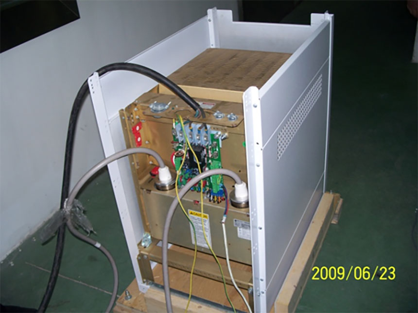
3) The main function of the high-voltage generator is to supply DC high voltage to the cathode and anode of the X-ray tube and the filament heating voltage. It is mainly composed of a high-voltage transformer, a filament transformer, a high-voltage rectifier, a high-voltage switching gate, transformer oil and a box that encapsulates the above components. After the high-voltage primary voltage is boosted by a high-voltage transformer and rectified by a high-voltage rectifier, it becomes a DC high voltage, which is added to the anode and cathode of the X-ray tube through a high-voltage cable. After the filament primary voltage is stepped down by the filament transformer, it is added to the X-ray tube filament through the cathode high-voltage cable.
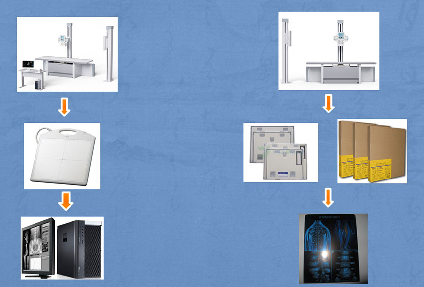
2. Imaging device
1) Traditional imaging devices: One type is a system that uses fluorescent screens (intensifying screens) and X-ray films as image carriers, operates in a darkroom, and doctors use viewing lamps for diagnosis; the other type is based on X-ray imaging devices such as image intensifiers and television systems. Radiography equipment, bright room operation, is the predecessor of digital imaging equipment.
2) Digital imaging device: Flat Panel Detector. The core technology of DR is the flat-panel detector. The flat-panel detector is a sophisticated and expensive device that plays a decisive role in imaging quality. Being familiar with the performance indicators of the detector can help us improve imaging quality and reduce X-ray radiation dose.
3) Display: a display device that reproduces the digital signals generated by the detector into images, including medical monitors (CRT), liquid crystal displays, and medical imaging liquid crystal displays (LCD).
3. Flat panel detector
Flat panel detectors are divided into amorphous selenium flat panel detectors (a-Se) and amorphous silicon flat panel detectors (a-Si)
Amorphous Selenium Flat Panel Detector (a-Se)
Amorphous selenium (a-Se) is a direct flat panel detector structure, mainly composed of a collector matrix, a selenium layer, a dielectric layer, a top electrode and a protective layer. The collector matrix consists of thin film transistors (TFTs) arranged in array elements. The amorphous selenium semiconductor material is vacuum evaporated on top of the thin film transistor array to form a thin film about 0.5 mm thick and 38 mm × 45 mm square. It is very sensitive to X-rays and has high image analysis capabilities.
Amorphous silicon flat panel detector (a-Si)
The amorphous silicon flat panel detector uses indirect digital X-ray imaging. Its basic structure is a layer of scintillator material (cesium iodide or gadolinium oxysulfide) on the surface, and the next layer is a photodiode circuit made of amorphous silicon. The bottom layer is the charge readout circuit.
The main models recommended by ysenmed are
YSFPD-V1717X wired board,
YSFPD-M1717V/
YSFPD-M1417V wireless board
3. Classification of medical X-ray machines
Classification of X-rays
1) According to working frequency: power frequency 50-60HZ / medium frequency / high frequency (20KHZ)
2) Classification by function: filming/fluoroscopy/filming+fluoroscopy
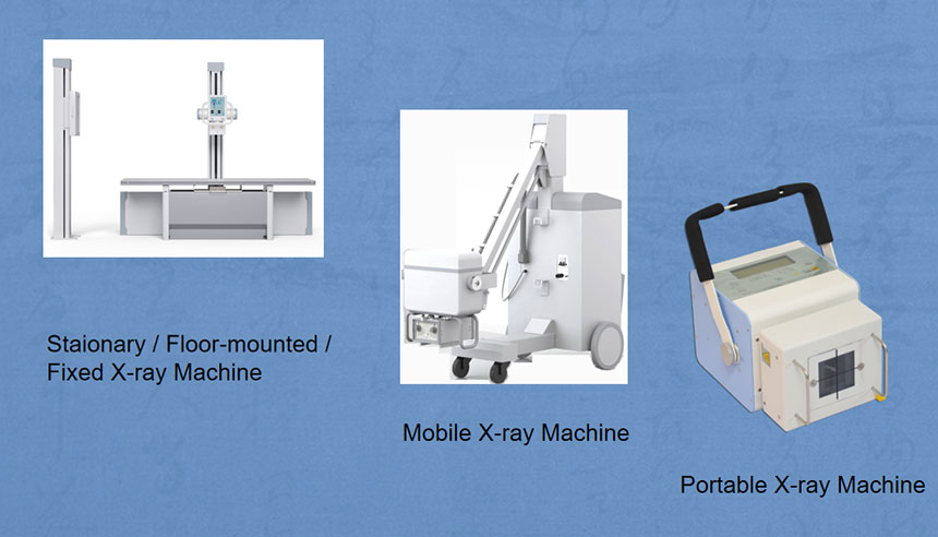
3) According to combination: mobile/fixed/portable
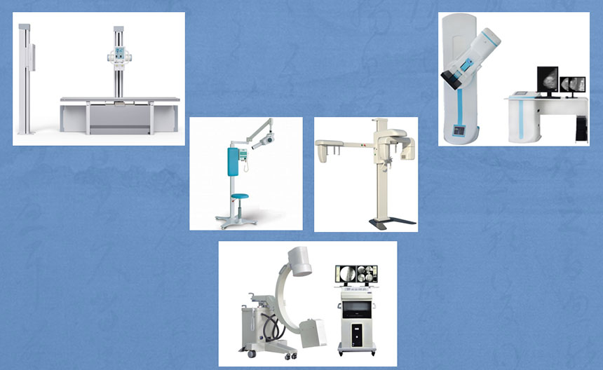
4) According to professional use: general purpose/dental machine/breast machine/C-arm
5) Press imaging mode: analog machine/digital X-ray machine
Power frequency 50-60HZ/medium frequency/high frequency (20KHZ)
Here are two of our best-selling machines recommended to you:
YSX100/YSX200/YSX300
Advantages: cheap
Disadvantages: Larger radiation, images are not as clear as high-frequency machines
YSX200G/YSX500G/YSX200GM YSX100GM
Advantages: less radiation and clearer images.
Disadvantages: The price is relatively high
Classification by function: Filming/fluoroscopy/filming+fluoroscopy
By combination: fixed/mobile/portable
By professional use: general purpose/dental machine/breast machine/C-arm
By imaging mode: Analog machine/Digital X-ray machine
YSENMED / Yuesen Med is a professional medical equipment company from China, compounded with the R&D and sale of medical device, is a leading supplier of medical equipment, especially in the field of medical x-ray machine.
Headquartered in Guangzhou, YSENMED covers the business of medical x-ray machines, x-ray accessories,ultrasound scanners, hospital sterilizers, anesthesia machines, ventilators, clinical lab equipment, operation room equipment, hospital furniture, and other medical equipment. Based on mutual trust & good service, our mission is to provide the most cost-effective medical equipment for hospital around the world.
In 2006, we exported 125 sets of medical x-ray machine to Congo at one time, setting a record of exporting medicalx-ray machine to foreign countries. In 2008, the same customer ordered another 150 x-ray units and 800 sets of surgical instruments. So far, we have established good cooperation with customers from 88 countries globally.
The latest x hot-selling recommendations in 2023
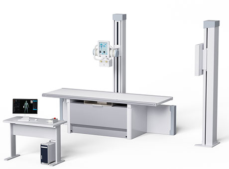
Simple bed model
Features:
1. There are 20kW, 32kW, 50kW, 65kW, etc. to choose from;
2. Simple operation;
3. Low price;
Analog model: YSX200-B1/YSX320-B1/YSX500-B1/YSX650-B1
Digital model: YSDR200-B1/YSDR320-B1/YSDR500-B1/YSDR650-B1
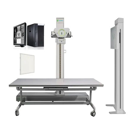
Deluxe bed
Features:
1. There are 20kW, 32kW, 50kW, 65kW, etc. to choose from;
2. The 10.4-inch large touch screen controls the movement of the column and tube;
3. High cost performance;
Analog model: YSX200G/YSX320G/YSX500G/YSX650G
Digital model: YSX200D/YSX320D/YSX500D/YSX650D
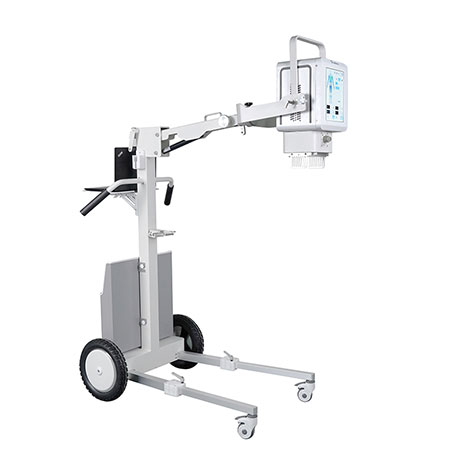
5.6KW high frequency portable machine
Mobile
Features:
1. It can be flexibly moved and is suitable for use in zoos, battlefields, and hospital beds;
2. The machine head can move up and down more than 120 degrees and rotate 360 degrees left and right;
3. The height can be flexibly adjusted and can be raised to a maximum height of 2.25 meters.
4. 5.6KW power, suitable for use within a body thickness of 25 cm
Features:
1. The machine head and the cassette can move synchronously, making it suitable for use in animal hospitals;
2. Built-in cassette tray, easy to upgrade DR;
3. Touch screen operation, simple and clear to use;
4. Three exposure methods: remote control, handbrake and touch screen exposure.
5. The combined machine head has a power of 5.6KW.
20/32KW veterinary X-ray machine
Features:
1. 20KW, 200mA, the shortest exposure time is 0.005S; 32KW, 400mA, the shortest exposure time is 0.005S;
2. After the bed is disassembled, it occupies a small space and is easy to install. It is suitable for domestic and foreign sales;
3. 1mAs step, 220 power supply stable output, unique in the market;
FAQ
Q:Is it for small clinic or large hospital?
A:What’s your requirment on power?
Q:Would you like floor-mounted or mobile type?
A:Which body parts do you mainly apply?


 By professional use: general purpose/dental machine/breast machine/C-arm
 By imaging mode: Analog machine/Digital X-ray machine


