Digital Subtraction Angiography (DSA) is a revolutionary imaging technique in the field of medical radiology that enables detailed visualization of blood vessels. It plays a crucial role in diagnosing various vascular conditions and guiding interventional procedures. DSA is commonly used in cardiology, neurology, and interventional radiology to examine the blood vessels of the heart, brain, and other organs. This article aims to provide a comprehensive overview of DSA, covering its definition, working principles, applications, advantages, operational procedure, and important considerations for its use.
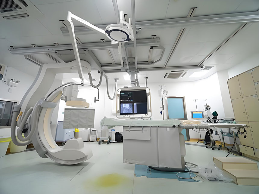
What is Digital Subtraction Angiography (DSA)?
Digital Subtraction Angiography (DSA) is an advanced medical imaging technique that uses digital image processing to enhance the visibility of blood vessels. The method combines the use of a contrast agent, which is injected into the blood vessels, and a sophisticated imaging system to capture detailed angiographic images. DSA involves "subtraction" techniques that remove the background anatomy (such as bone and soft tissue) from the images, leaving only the blood vessels for clear visualization.
Unlike traditional X-ray angiography, which can show both the blood vessels and surrounding tissues in a single image, DSA is able to subtract the background and isolate the vascular structures, providing high-resolution images of blood flow. This subtraction of background allows for superior contrast and more accurate detection of abnormalities in the vascular system.
How Does Digital Subtraction Angiography (DSA) Work?
DSA works by utilizing a series of X-ray images taken before and after the injection of a contrast dye into the patient's blood vessels. Here's a breakdown of the process:
- Injection of Contrast Agent: A radiopaque dye is injected into the blood vessels, making the vessels visible on X-ray images.
- Image Acquisition: A series of X-ray images are taken at various angles, both before and after the contrast agent is introduced.
- Subtraction Process: The computer system then subtracts the initial images (without contrast) from the subsequent images (with contrast). This process removes the non-vascular structures, leaving behind only the blood vessels in the final image.
- Enhanced Visualization: The resulting image highlights any abnormalities or blockages in the blood vessels, allowing for precise diagnosis and treatment planning.
The subtraction process is what makes DSA stand out from traditional angiography techniques. By removing the non-essential structures, DSA provides an incredibly clear and detailed view of the vascular anatomy, which is essential for accurate diagnosis and effective treatment.
Applications of Digital Subtraction Angiography (DSA)
DSA is widely used in clinical practice due to its ability to provide detailed imaging of blood vessels, making it invaluable in various medical specialties. Some common applications of DSA include:
-
Cardiovascular Imaging:
- Coronary Artery Disease (CAD): DSA is a critical tool in diagnosing CAD by visualizing blockages or narrowing of the coronary arteries.
- Peripheral Artery Disease (PAD): It helps identify blockages or stenosis in peripheral blood vessels, particularly in the legs.
- Aneurysms: DSA is used to detect aneurysms, particularly in the brain (cerebral aneurysms) or the aorta.
-
Neurological Imaging:
- Brain Imaging: DSA plays a crucial role in diagnosing cerebrovascular diseases such as stroke, aneurysms, and arteriovenous malformations (AVMs).
- Cerebral Angiography: DSA is essential for evaluating the blood supply to the brain and detecting abnormalities such as cerebral hemorrhages or ischemia.
-
Interventional Radiology:
- DSA is used as a guide for a variety of interventional procedures, such as stent placement, embolization (blocking blood flow to certain areas), and catheter-based treatments.
- Endovenous Laser Treatment (EVLT) for varicose veins relies on DSA for precision.
-
Oncology:
- DSA can be used for tumor embolization, where blood supply to tumors is blocked to shrink the tumors or prevent them from growing.
-
Renal Imaging:
- DSA is used to evaluate the renal vasculature, particularly in the case of renal artery stenosis, which can lead to hypertension.
Advantages of Digital Subtraction Angiography (DSA)
There are numerous benefits of DSA, making it an indispensable tool in modern medical practice:
-
High-Resolution Imaging: DSA provides extremely detailed images of blood vessels, which are vital for the accurate diagnosis of vascular conditions such as blockages, aneurysms, and other abnormalities.
-
Enhanced Precision for Interventions: The detailed images produced by DSA help guide interventional procedures, such as stenting or embolization, with high precision. This reduces the risk of complications and improves patient outcomes.
-
Real-Time Imaging: One of the major advantages of DSA is that it provides real-time images, which is particularly useful during interventional procedures. Doctors can see live images of the vessels, ensuring that the procedure is being carried out correctly.
-
Minimal Patient Discomfort: DSA is a non-invasive procedure, meaning that patients do not need to undergo surgery to assess their vascular condition. The process only involves the injection of a contrast agent, which is typically well-tolerated.
-
Reduced Need for Multiple Procedures: The high accuracy of DSA means that fewer follow-up procedures are often needed. Additionally, it can replace more invasive surgical procedures in some cases, thus reducing the overall risk to the patient.
-
Multipurpose Imaging: DSA can be used for a wide variety of medical conditions, from cardiovascular issues to neurological diseases, making it a versatile diagnostic tool.
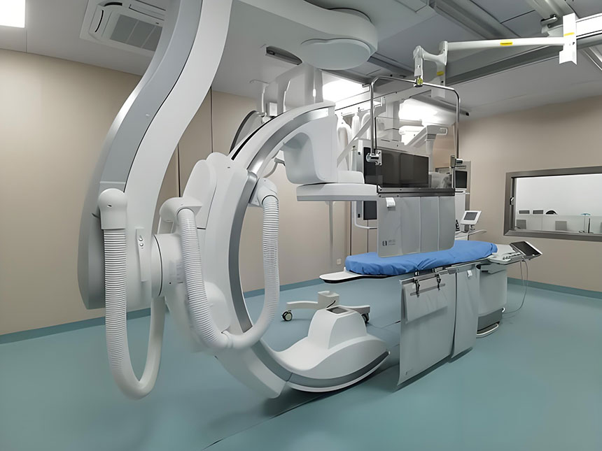
How DSA is Performed: Step-by-Step Guide
The procedure for performing Digital Subtraction Angiography involves several key steps. Here's what patients can expect during the procedure:
- Preparation: The patient is positioned on an examination table, and an intravenous line is established for the contrast injection.
- Local Anesthesia: Depending on the procedure, local anesthesia may be applied at the injection site (typically the groin, arm, or neck).
- Contrast Injection: A contrast agent is injected into the blood vessels to highlight them during the imaging process.
- Image Acquisition: A series of X-ray images is taken, both before and after the contrast is injected.
- Image Processing: The images are processed by a computer system that subtracts the non-vascular structures to produce clear images of the blood vessels.
- Analysis: A radiologist or specialist analyzes the images to detect any abnormalities or issues in the vascular system.
Important Considerations and Risks
While DSA is a powerful diagnostic tool, it is important to consider the potential risks and precautions:
-
Contrast Agent Reactions: Some patients may experience allergic reactions to the contrast dye. It is important for patients to inform their doctor about any known allergies before the procedure.
-
Radiation Exposure: Like other X-ray-based imaging techniques, DSA exposes patients to a small amount of radiation. Although the risks are minimal, it is important to use this technique judiciously.
-
Kidney Function: Since the contrast agent is processed by the kidneys, patients with pre-existing kidney issues should be carefully evaluated before undergoing DSA.
-
Infection Risks: There is a slight risk of infection at the site where the catheter is inserted, but this is rare.
-
Post-Procedure Care: Patients may be monitored for a short period after the procedure to ensure there are no complications, especially if they are undergoing an interventional procedure.
Conclusion
Digital Subtraction Angiography (DSA) is a crucial imaging technique that enables the detailed visualization of blood vessels, offering significant advantages in diagnosing and treating a variety of vascular conditions. With its ability to produce high-resolution, real-time images, DSA is indispensable in interventional procedures and provides critical insight into the health of the vascular system. However, as with any medical procedure, it is important to be aware of the risks and to discuss these with a healthcare professional. By understanding the principles, applications, and advantages of DSA, patients can make informed decisions about their healthcare options.