In the world of medical imaging, the advent of digital technologies has transformed diagnostic practices. Among the most significant advancements is the development and use of Flat Panel Detectors (FPD) in radiology. FPDs are essential components in modern imaging systems, especially for digital radiography (DR), mammography, and fluoroscopy. Their ability to provide high-resolution, fast imaging with low radiation exposure has made them a staple in healthcare settings worldwide.
This comprehensive guide aims to explain what Flat Panel Detectors are, how they work, their applications, advantages, operational processes, and important considerations when using them.

What is a Flat Panel Detector (FPD)?
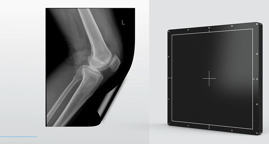
A Flat Panel Detector (FPD) is a type of image receptor used in digital radiography (DR) systems to capture X-ray images and convert them into digital data. Unlike traditional film-based imaging, FPDs utilize advanced electronic technology to produce high-quality images without the need for film or a traditional photochemical process.
FPDs are integral to various medical imaging modalities such as general X-ray imaging, mammography, fluoroscopy, and even computed tomography (CT) systems. They come in two main types: direct and indirect detectors.
-
Direct FPDs: These detectors use a photosensitive material like selenium to directly convert X-rays into electrical signals. This process eliminates the need for any intermediate steps.
-
Indirect FPDs: These detectors first convert X-rays into light using a scintillation material (like cesium iodide), and then the light is transformed into an electrical signal via a photodiode.
How Do Flat Panel Detectors Work?
Flat Panel Detectors function by capturing X-rays and converting them into digital signals. Here’s how the process works:
-
X-ray Exposure: The patient is exposed to X-rays, and the FPD is positioned to detect the radiation passing through the body. The X-rays interact with the detector and are absorbed by the active layer.
-
Conversion: In direct FPDs, the X-rays interact with a photoconductive layer (such as selenium), which converts the radiation into an electrical charge. In indirect FPDs, the X-rays are first converted into light by a scintillator material (like cesium iodide), and the light is then detected and converted into an electrical signal by a photodiode.
-
Digital Signal Processing: The electrical signal generated by the detector is transmitted to a computer where it is converted into a digital image. The image can then be manipulated, analyzed, and stored in a digital format.
-
Image Display: The final digital image is displayed on a monitor for immediate review by the radiologist or physician, allowing for quicker diagnosis and treatment planning.
Applications of Flat Panel Detectors
Flat Panel Detectors are versatile tools used across various medical imaging applications. Their ability to provide real-time, high-quality images with low radiation exposure makes them indispensable in modern healthcare. Below are some common applications:
-
General Radiography: FPDs are widely used in routine X-ray examinations, such as chest X-rays, bone scans, and abdominal imaging. They provide clear, high-resolution images with reduced exposure to radiation compared to traditional film-based systems.
-
Mammography: FPDs are crucial in digital mammography, where high-resolution images of the breast tissue are necessary for detecting abnormalities such as lumps or microcalcifications. They provide excellent contrast and resolution, which is essential for early breast cancer detection.
-
Fluoroscopy: In fluoroscopic procedures, FPDs are used to capture real-time images of dynamic processes, such as the movement of a contrast medium through the digestive or cardiovascular system. These detectors enable continuous imaging, which is vital for guiding procedures like angiography and catheter placement.
-
Interventional Radiology: FPDs are widely used in interventional radiology for procedures such as catheter placement, embolization, and stent insertion. Their ability to provide high-resolution images in real-time is critical for the precision required in these minimally invasive procedures.
-
Computed Tomography (CT): In some CT systems, FPDs are used to detect X-ray radiation and generate detailed cross-sectional images of the body. They provide better image quality and lower radiation exposure compared to traditional CT detectors.
-
Veterinary Imaging: FPDs are also used in veterinary medicine for imaging animals, where high-resolution and real-time imaging are equally important.
Advantages of Flat Panel Detectors
Flat Panel Detectors offer several advantages over traditional film-based systems, making them a preferred choice in modern medical imaging. Some of the key benefits include:
-
High-Resolution Imaging: FPDs provide exceptional image quality with greater resolution and contrast. This is particularly important in detecting small or subtle abnormalities, such as tumors, fractures, or vascular irregularities.
-
Reduced Radiation Exposure: One of the most significant benefits of FPDs is their ability to capture high-quality images with lower radiation doses. This reduction in radiation exposure is crucial for patient safety, especially for vulnerable populations such as children and pregnant women.
-
Fast Image Acquisition: FPDs enable immediate image acquisition and display, eliminating the need for film development and providing real-time imaging. This allows for quicker diagnosis and faster decision-making in critical situations.
-
Digital Image Storage and Sharing: The digital nature of FPD images means that they can be stored electronically, eliminating the need for physical film storage. Digital images can also be easily shared between healthcare facilities, aiding in remote consultations and second opinions.
-
Enhanced Post-Processing: Digital images obtained through FPDs can be manipulated and processed to enhance image clarity, contrast, and sharpness. This post-processing capability improves diagnostic accuracy and allows radiologists to view images from different perspectives.
-
Lower Operating Costs: Unlike traditional film-based systems that require film, chemicals, and physical storage space, FPDs are more cost-effective in the long run due to the elimination of these consumables. Additionally, FPDs require less maintenance and fewer physical resources.
How to Operate Flat Panel Detectors
Operating a Flat Panel Detector is relatively straightforward, but it requires a basic understanding of the system and the imaging procedure. Below are the general steps involved in operating FPDs:
-
Preparation: Ensure the patient is properly positioned on the imaging table. The radiologist or technician will also need to select the appropriate settings on the imaging machine, including exposure factors (such as kilovoltage and milliampere settings).
-
X-ray Exposure: The FPD will detect the X-ray exposure passing through the patient’s body. The detector automatically captures the X-rays, and the images are processed and displayed digitally.
-
Image Processing: Once the X-ray exposure is complete, the FPD system processes the data and generates a digital image. This image can be further manipulated or enhanced using software for better visualization.
-
Review and Diagnosis: The final image is reviewed on a digital display monitor by a radiologist or healthcare provider to assess the patient’s condition.
-
Post-Examination: After the imaging procedure, the images are stored digitally in the hospital’s imaging system, allowing for easy access, sharing, and long-term storage.
Important Considerations When Using Flat Panel Detectors
Although Flat Panel Detectors are highly effective and safe for medical imaging, there are certain considerations that healthcare providers should keep in mind:
-
Image Artifacts: Just like any other imaging technology, FPDs can sometimes produce artifacts—unwanted imperfections in the images. These can result from improper patient positioning, motion, or technical malfunctions. It is essential to ensure proper setup and calibration of the system to avoid such artifacts.
-
Calibration: FPD systems require periodic calibration to ensure optimal performance. Regular calibration ensures that the system produces accurate and high-quality images.
-
Radiation Protection: While FPDs reduce radiation exposure compared to traditional film-based systems, proper radiation safety protocols should still be followed. Protective shields, proper positioning, and limiting exposure time should always be observed to minimize any risks associated with radiation.
-
Maintenance: FPDs are complex devices that require regular maintenance to function at their best. Regular servicing and checks by trained professionals ensure the longevity and efficiency of the equipment.
-
Cost: While FPDs are more cost-effective in the long term, the initial investment can be high. Hospitals and clinics must weigh the upfront cost against the long-term benefits of enhanced image quality and reduced operational costs.

Conclusion
Flat Panel Detectors (FPD) represent a significant advancement in medical imaging technology. Their ability to provide high-resolution images with reduced radiation exposure makes them a cornerstone of modern diagnostic practices in radiology, cardiology, and oncology. With applications ranging from general radiography to specialized procedures like mammography and fluoroscopy, FPDs have revolutionized the way healthcare professionals approach diagnosis and treatment.
Their advantages, including fast imaging, digital storage, and enhanced image quality, make FPDs an essential tool for any modern healthcare facility. As technology continues to evolve, we can expect further innovations that will continue to improve diagnostic accuracy, patient care, and overall healthcare outcomes.


